Anatomy and Physiology Lab Review Sheet 36 Answer
The trillions of cells in the body require an abundant and continuous supply of oxygen to comport out their vital functions. We cannot "practise without oxygen" for even a little while, as nosotros tin without nutrient or water.
Functions of the Respiratory System
The functions of the respiratory arrangement are:
- Oxygen supplier.The job of the respiratory organization is to keep the trunk constantly supplied with oxygen.
- Elimination.Elimination of carbon dioxide.
- Gas exchange.The respiratory system organs oversee the gas exchanges that occur between the blood and the external surroundings.
- Passageway.Passageways that allow air to achieve the lungs.
- Humidifier.Purify, humidify, and warm incoming air.
Anatomy of the Respiratory Organization
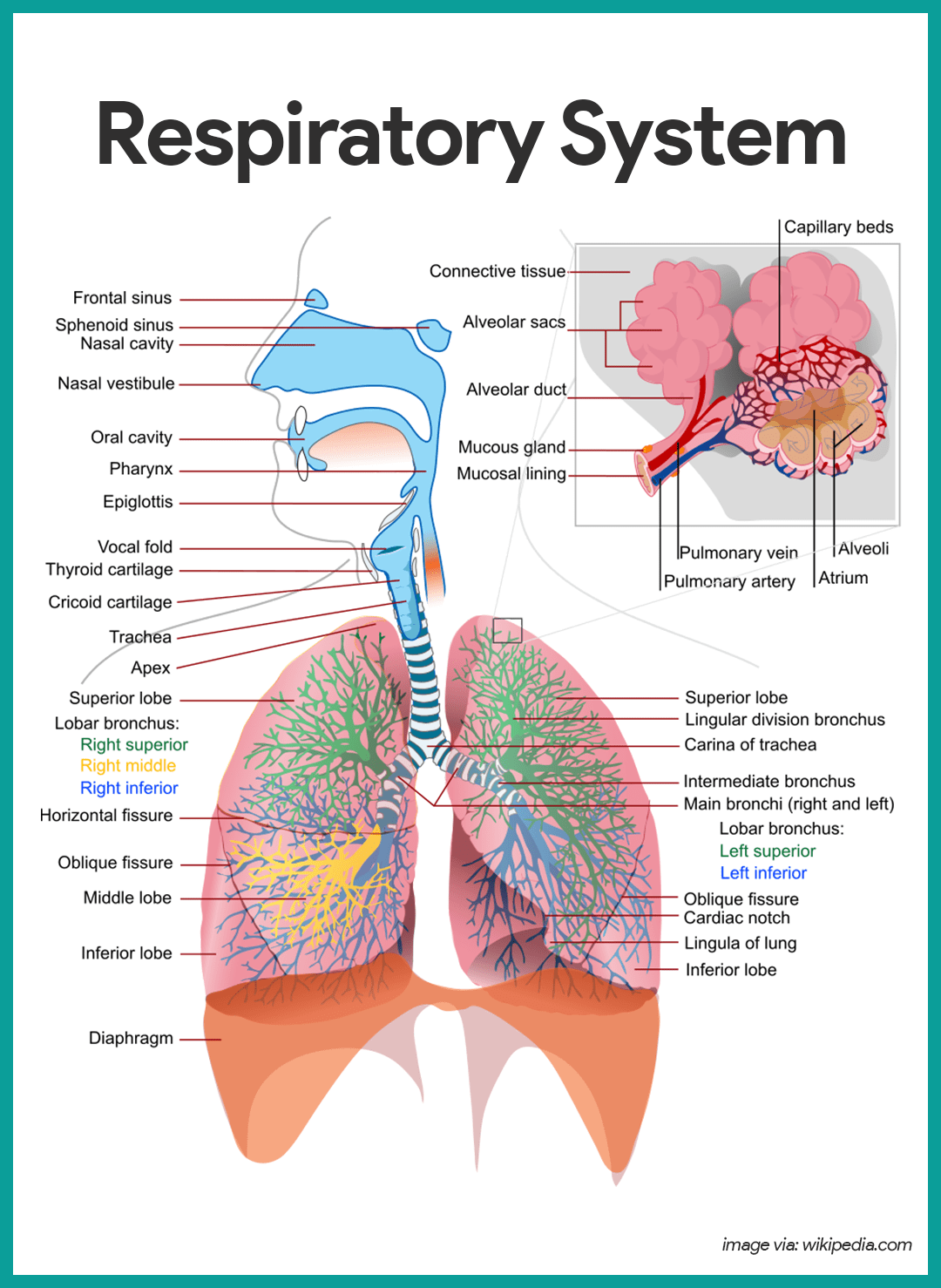
The organs of the respiratory system include the nose, pharynx, larynx, trachea, bronchi, and their smaller branches, and the lungs, which contain the alveoli.
The Olfactory organ
The olfactory organ is the just externally visible part of the respiratory system.
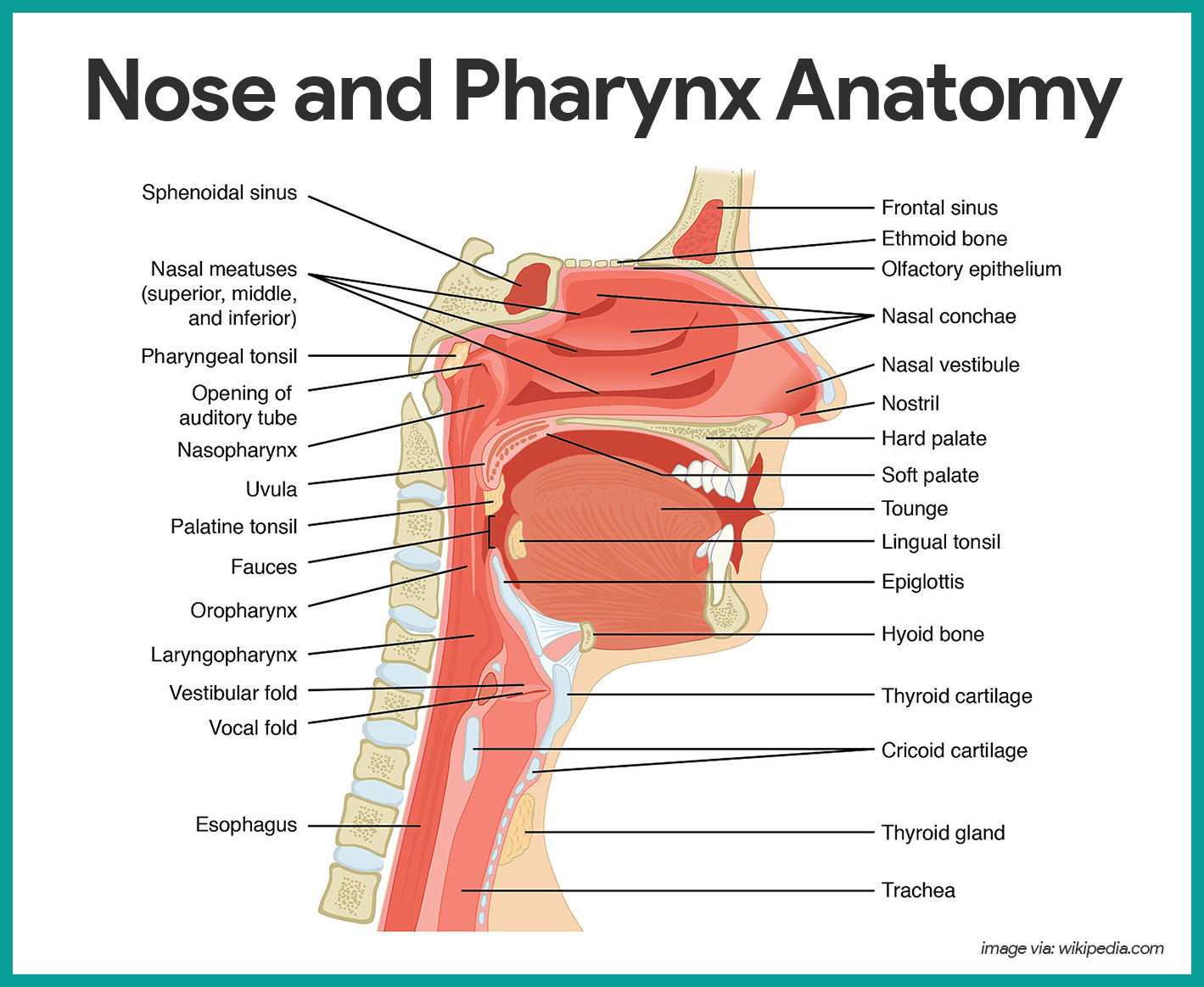
- Nostrils.During breathing, air enters the nose by passing through the nostrils, or nares.
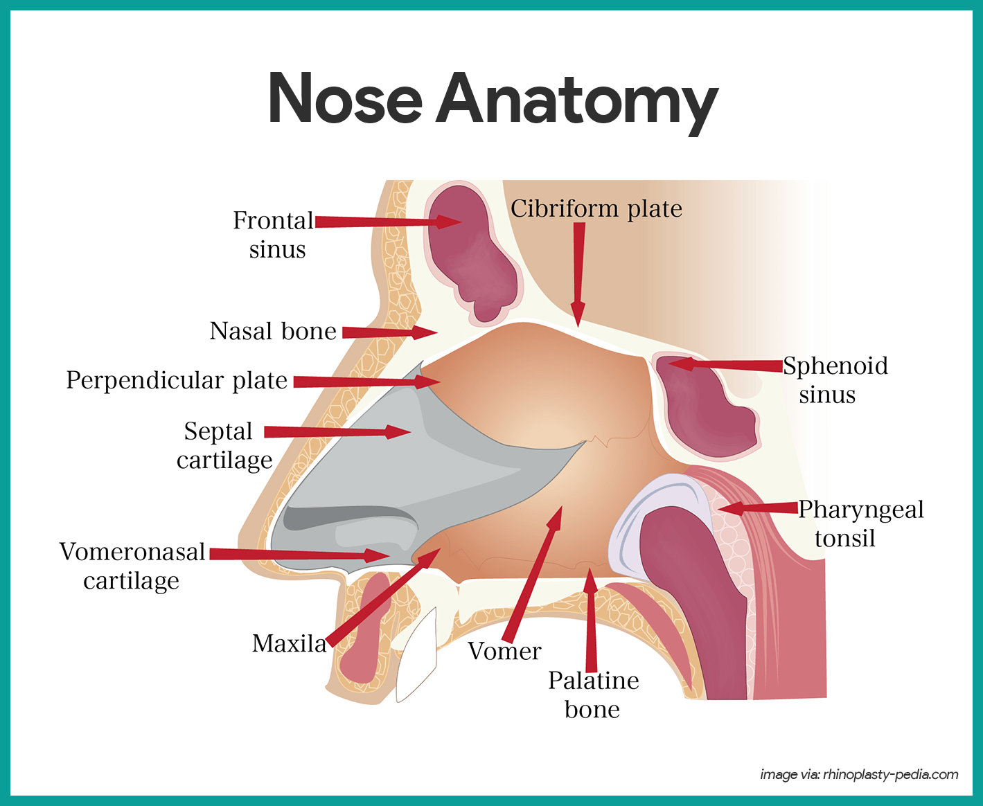
- Nasal cavity. The interior of the olfactory organ consists of the nasal cavity, divided by a midline nasal septum.
- Olfactory receptors. The olfactory receptors for the sense of smell are located in the mucosa in the slitlike superior function of the nasal cavity, just beneath the ethmoid os.
- Respiratory mucosa. The rest of the mucosal lining, the nasal crenel called the respiratory mucosa, rests on a rich network of thin-walled veins that warms the air as information technology flows past.
- Fungus.In addition, the sticky mucus produced by the mucosa's glands moistens the air and traps incoming bacteria and other foreign droppings, and lysozyme enzymes in the fungus destroy leaner chemically.
- Ciliated cells. The ciliated cells of the nasal mucosa create a gentle current that moves the sail of contaminated mucus posteriorly toward the throat, where it is swallowed and digested by breadbasket juices.
- Conchae.The lateral walls of the nasal cavity are uneven owing to three mucosa-covered projections, or lobes called conchae, which greatly increase the surface area of the mucosa exposed to the air, and also increase the air turbulence in the nasal cavity.
- Palate. The nasal cavity is separated from the oral cavity below past a sectionalisation, the palate; anteriorly, where the palate is supported by bone, is the hard palate; the unsupported posterior part is the soft palate.
- Paranasal sinuses. The nasal cavity is surrounded by a band of paranasal sinuses located in the frontal, sphenoid, ethmoid, and maxillary basic; theses sinuses lighten the skull, and they act as a resonance sleeping accommodation for speech.
Pharynx
- Size. The pharynx is a muscular passageway about 13 cm (5 inches) long that vaguely resembles a short length of red garden hose.
- Function.Commonly called the throat, the pharynx serves as a mutual passageway for food and air.
- Portions of the pharynx. Air enters the superior portion, the nasopharynx, from the nasal cavity and so descends through the oropharynx and laryngopharynx to enter the larynx beneath.
- Pharyngotympanic tube. The pharyngotympanic tubes, which drain the middle ear open up into the nasopharynx.
- Pharyngeal tonsil. The pharyngeal tonsil, often called adenoid is located high in the nasopharynx.
- Palatine tonsils. The palatine tonsils are in the oropharynx at the end of the soft palate.
- Lingual tonsils. The lingual tonsils lie at the base of operations of the tongue.
Larynx
The larynx or vocalization box routes air and food into the proper channels and plays a role in speech.
- Structure.Located junior to the throat, it is formed by eight rigid hyaline cartilages and a spoon-shaped flap of rubberband cartilage, the epiglottis.
- Thyroid cartilage. The largest of the hyaline cartilages is the shield-shaped thyroid cartilage, which protrudes anteriorly and is commonly called Adam's apple.
- Epiglottis.Sometimes referred to every bit the "guardian of the airways", the epiglottis protects the superior opening of the larynx.
- Vocal folds. Part of the mucous membrane of the larynx forms a pair of folds, called the vocal folds, or true song cords, which vibrate with expelled air and allows u.s. to speak.
- Glottis.The slitlike passageway betwixt the vocal folds is the glottis.
Trachea
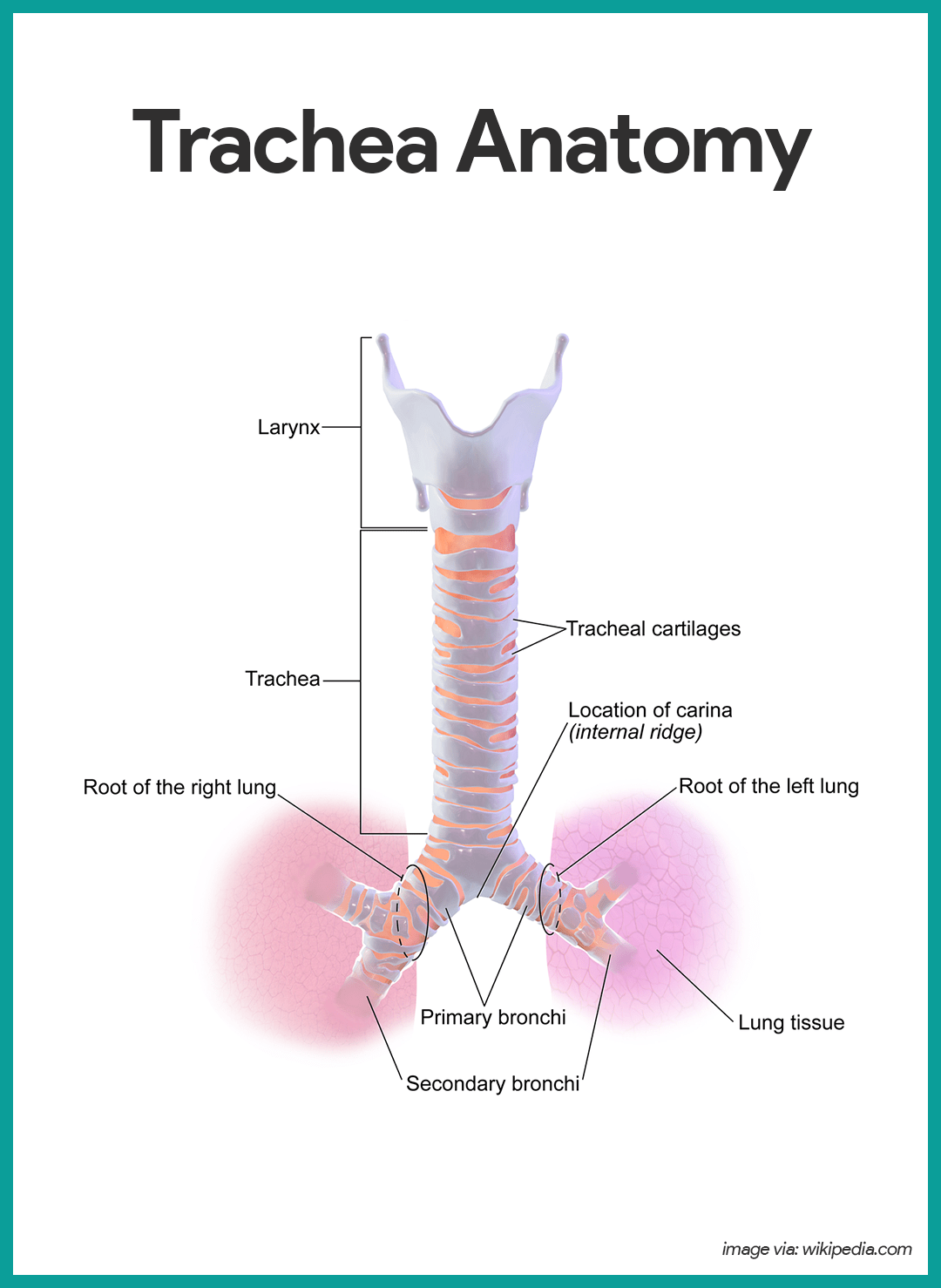
- Length.Air entering the trachea or windpipe from the larynx travels downwardly its length (10 to 12 cm or about 4 inches) to the level of the 5th thoracic vertebra, which is approximately midchest.
- Structure.The trachea is adequately rigid because its walls are reinforced with C-shaped rings of hyaline cartilage; the open parts of the rings abut the esophagus and let information technology to expand anteriorly when we swallow a large piece of nutrient, while the solid portions support the trachea walls and keep it patent, or open, in spite of the pressure changes that occur during breathing.
- Cilia.The trachea is lined with ciliated mucosa that crush continuously and in a direction reverse to that of the incoming air as they propel fungus, loaded with dust particles and other droppings away from the lungs to the throat, where it tin can be swallowed or spat out.
Main Bronchi
- Structure.The right and left main (chief) bronchi are formed by the partitioning of the trachea.
- Location.Each chief bronchus runs obliquely earlier it plunges into the medial depression of the lung on its own side.
- Size.The correct main bronchus is wider, shorter, and straighter than the left.
Lungs
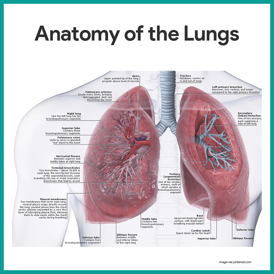
- Location.The lungs occupy the unabridged thoracic cavity except for the most central area, the mediastinum, which houses the middle, the bully claret vessels, bronchi, esophagus, and other organs.
- Apex.The narrow, superior portion of each lung, the apex, is but deep to the clavicle.
- Base.The wide lung surface area resting on the diaphragm is the base.
- Sectionalization.Each lung is divided into lobes by fissures; the left lung has two lobes, and the right lung has 3.
- Pleura.The surface of each lung is covered with a visceral serosa called the pulmonary, or visceral pleura and the walls of the thoracic cavity are lined by the parietal pleura.
- Pleural fluid. The pleural membranes produce pleural fluid, a glace serous secretion which allows the lungs to glide easily over the thorax wall during breathing movements and causes the two pleural layers to cling together.
- Pleural infinite. The lungs are held tightly to the thorax wall, and the pleural infinite is more of a potential space than an bodily one.
- Bronchioles.The smallest of the conducting passageways are the bronchioles.
- Alveoli.The terminal bronchioles lead to the respiratory zone structures, even smaller conduits that eventually terminate in alveoli, or air sacs.
- Respiratory zone. The respiratory zone, which includes the respiratory bronchioles, alveolar ducts, alveolar sacs, and alveoli, is the simply site of gas substitution.
- Conducting zone structures. All other respiratory passages are conducting zone structures that serve as conduits to and from the respiratory zone.
- Stroma.The balance of the lung tissue, its stroma, is mainly elastic connective tissue that allows the lungs to recoil passively as nosotros breathe.
The Respiratory Membrane
- Wall structure. The walls of the alveoli are composed largely of a single, thin layer of squamous epithelial cells.
- Alveolar pores. Alveolar pores connecting neighboring air sacs and provide alternative routes for air to achieve alveoli whose feeder bronchioles have been clogged by mucus or otherwise blocked.
- Respiratory membrane. Together, the alveolar and capillary walls, their fused basement membranes, and occasional elastic fibers construct the respiratory membrane (air-blood barrier), which has gas (air) flowing by on one side and blood flowing past on the other.
- Alveolar macrophages. Remarkably efficient alveolar macrophages sometimes called "grit cells", wander in and out of the alveoli picking up leaner, carbon particles, and other debris.
- Cuboidal cells. Also scattered amidst the epithelial cells that form most of the alveolar walls are chunky cuboidal cells, which produce a lipid (fatty) molecule called surfactant, which coats the gas-exposed alveolar surfaces and is very of import in lung function.
Physiology of the Respiratory System
The major function of the respiratory organization is to supply the body with oxygen and to dispose of carbon dioxide. To do this, at least 4 distinct events, collectively called respiration, must occur.
Respiration
- Pulmonary ventilation. Air must move into and out of the lungs then that gasses in the air sacs are continuously refreshed, and this procedure is commonly called breathing.
- External respiration. Gas exchange between the pulmonary blood and alveoli must take place.
- Respiratory gas transport. Oxygen and carbon dioxide must be transported to and from the lungs and tissue cells of the body via the bloodstream.
- Internal respiration. At systemic capillaries, gas exchanges must be fabricated betwixt the claret and tissue cells.
Mechanics of Breathing
- Rule.Book changes atomic number 82 to force per unit area changes, which pb to the flow of gasses to equalize pressure.
- Inspiration.Air is flowing into the lungs; chest is expanded laterally, the rib muzzle is elevated, and the diaphragm is depressed and flattened; lungs are stretched to the larger thoracic volume, causing the intrapulmonary pressure to fall and air to catamenia into the lungs.
- Expiration.Air is leaving the lungs; the chest is depressed and the lateral dimension is reduced, the rib muzzle is descended, and the diaphragm is elevated and dome-shaped; lungs recoil to a smaller volume, intrapulmonary pressure rises, and air flows out of the lung.
- Intrapulmonary volume. Intrapulmonary volume is the volume within the lungs.
- Intrapleural pressure. The normal pressure level within the pleural infinite, the intrapleural pressure, is always negative, and this is the major factor preventing the plummet of the lungs.
- Nonrespiratory air movements. Nonrespiratory movements are a result of reflex activity, but some may be produced voluntarily such as cough, sneeze, crying, laughing, hiccups, and yawn.
Respiratory Volumes and Capacities
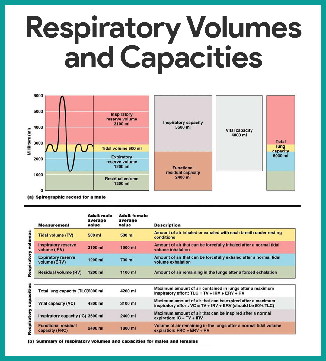
- Tidal book. Normal quiet breathing moves approximately 500 ml of air into and out of the lungs with each breath.
- Inspiratory reserve book. The amount of air that can be taken in forcibly over the tidal volume is the inspiratory reserve volume, which is commonly between 2100 ml to 3200 ml.
- Expiratory reserve volume. The corporeality of air that can be forcibly exhaled afterward a tidal expiration, the expiratory reserve volume, is approximately 1200 ml.
- Residual volume. Even after the virtually strenuous expiration, about 1200 ml of air yet remains in the lungs and information technology cannot be voluntarily expelled; this is called rest volume, and it is important considering it allows gas exchange to go on continuously even between breaths and helps to keep the alveoli inflated.
- Vital capacity. The total amount of exchangeable air is typically effectually 4800 ml in healthy young men, and this respiratory capacity is the vital chapters, which is the sum of the tidal volume, inspiratory reserve book, and the expiratory reserve book.
- Expressionless space volume. Much of the air that enters the respiratory tract remains in the conducting zone passageways and never reaches the alveoli; this is called the expressionless space volume and during a normal tidal jiff, it amounts to about 150 ml.
- Functional volume. The functional book, which is the air that really reaches the respiratory zone and contributes to gas exchange, is about 350 ml.
- Spirometer.Respiratory capacities are measured with a spirometer, wherein equally a person breathes, the volumes of air exhaled can be read on an indicator, which shows the changes in air volume inside the appliance.
Respiratory Sounds
- Bronchial sounds. Bronchial sounds are produced by air rushing through the large respiratory passageways (trachea and bronchi).
- Vesicular animate sounds. Vesicular animate sounds occur as air fills the alveoli, and they are soft and resemble a muffled breeze.
External Respiration, Gas Transport, and Internal Respiration
- External respiration. External respiration or pulmonary gas commutation involves the oxygen being loaded and carbon dioxide existence unloaded from the blood.
- Internal respiration. In internal respiration or systemic capillary gas substitution, oxygen is unloaded and carbon dioxide is loaded into the claret.
- Gas transport. Oxygen is transported in the blood in ii ways: most attaches to hemoglobin molecules within the RBCs to course oxyhemoglobin, or a very pocket-size amount of oxygen is carried dissolved in the plasma; while carbon dioxide is transported in plasma every bit bicarbonate ion, or a smaller amount (betwixt xx to 30 percent of the transported carbon dioxide) is carried inside the RBCs jump to hemoglobin.
Control of Respiration
Neural Regulation
- Phrenic and intercostal nerves. These two nerves regulate the activity of the respiratory muscles, the diaphragm, and external intercostals.
- Medulla and pons. Neural centers that control respiratory rhythm and depth are located mainly in the medulla and pons; the medulla, which sets the bones rhythm of breathing, contains a pacemaker, or cocky-exciting inspiratory heart, and an expiratory eye that inhibits the pacemaker in a rhythmic way; pons centers appear to smooth out the bones rhythm of inspiration and expiration set by the medulla.
- Eupnea.The normal respiratory rate is referred to every bit eupnea, and it is maintained at a rate of 12 to xv respirations/minute.
- Hyperpnea.During practice, we breathe more vigorously and deeply because the brain centers transport more impulses to the respiratory muscles, and this respiratory blueprint is chosen hyperpnea.
Non-neural Factors Influencing Respiratory Charge per unit and Depth
- Physical factors. Although the medulla's respiratory centers set the basic rhythm of breathing, in that location is no question that concrete factors such as talking, coughing, and exercising tin can modify both the charge per unit and depth of breathing, equally well as an increased torso temperature, which increases the charge per unit of breathing.
- Volition (conscious control). Voluntary control of breathing is limited, and the respiratory centers will merely ignore messages from the cortex (our wishes) when the oxygen supply in the blood is getting low or blood pH is falling.
- Emotional factors. Emotional factors as well change the rate and depth of animate through reflexes initiated by emotional stimuli acting through centers in the hypothalamus.
- Chemical factors. The nearly important factors that alter respiratory rate and depth are chemical- the levels of carbon dioxide and oxygen in the blood; increased levels of carbon dioxide and decreased blood pH are the nigh of import stimuli leading to an increase in the rate and depth of breathing, while a subtract in oxygen levels become important stimuli when the levels are dangerously low.
- Hyperventilation.Hyperventilation blows off more carbon dioxide and decreases the amount of carbonic acid, which returns blood pH to normal range when carbon dioxide or other sources of acids begin to accumulate in the blood.
- Hypoventilation.Hypoventilation or extremely ho-hum or shallow breathing allows carbon dioxide to accumulate in the blood and brings claret pH back into normal range when blood starts to become slightly alkaline.
Exercise Quiz: Respiratory Organisation Anatomy and Physiology
Here's a 10-particular quiz nearly the written report guide. Please visit our nursing test bank page for more than NCLEX practice questions.
1. Which of the following descriptions regarding the larynx is CORRECT?
A. The almost inferior cartilage in the larynx is the epiglottis.
B. Unlike the other cartilages of the larynx, the epiglottis consists of hyaline cartilage.
C. The larynx contains 4 unpaired cartilages.
D. When the vestibular folds come together, they prevent air from leaving the lungs.
1. Respond: D. When the vestibular folds come together, they prevent air from leaving the lungs.
D: When the vestibular folds come together, they prevent air from leaving the lungs, such as when a person holds his breath. Along with the epiglottis, the vestibular folds also prevent nutrient and liquids from entering the larynx.
A: The virtually inferior cartilage of the larynx is the unpaired cricoid cartilage, which forms the base of the larynx on which the other cartilages residue.
B: The epiglottis differs from the other cartilages in that it consists of elastic cartilage rather than hyaline cartilage.
C: The larynx consists of an outer casing of nine cartilages that are connected to one another by muscles and ligaments. Three of the nine cartilages are unpaired, and six of them course iii pairs.
2. Given these respiratory passageways:
1. alveoli
two. bronchi
iii. bronchioles
4. respiratory bronchioles
5. terminal bronchioles
From largest to smallest, the authentic order for these passageways is:
A. two, 4, five, 3, 1
B. 2, iv, iii, five, one
C. 2, three, 5, 4, 1
D. 2, 3, four, v, ane
2. Answer: C. 2, three, 5, 4, 1
The primary bronchi branch many times to class the tracheobronchial tree. Within the lungs, the main airways (bronchi) co-operative off into smaller and smaller passageways. The conducting portion is made up of nasal cavities, nasopharynx, larynx, trachea, bronchi and bronchioles. The trachea branches to requite rise to two primary (main) bronchi. These and so branch successively to requite rising in plough to secondary and 3rd bronchi. These and so branch to give rise to several orders of progressively smaller airways called bronchioles, the smallest of which are called terminal bronchioles. These are the last components of the conducting portion of the respiratory system. Terminal bronchioles requite ascent to respiratory bronchioles, which ultimately lead to the alveoli.
three. The right lung has ___ lobes and ___ bronchopulmonary segments.
A. 2, 9
B. 2, 10
C. 3, 9
D. iii, x
3. Reply: D. 3, 10
The correct lung has three lobes called the superior, middle, and inferior lobes. The left lung has two lobes called the superior and inferior lobes. Each lobe is divided into bronchopulmonary segments. There are 9 bronchopulmonary segments in the left lung and x in the right lung.
4. The pleura that covers the surface of the lungs is the:
A. Pleural Cavity
B. Pleural Fluid
C. Visceral Pleura
D. Parietal Pleura
4. Answer: C. Visceral Pleura
C: The visceral pleura covers the surface of the lung.
A,B: The pleural cavity, between the parietal and visceral pleurae, is filled with a small-scale volume of pleural fluid produced past the pleural membranes.
D: The parietal pleura lines the walls of the thorax, diaphragm, and mediastinum.
5. The muscles of inspiration include the diaphragm and internal intercostal muscles. The argument is:
A. True
B. False
C. Partially true
D. Partially false
5. Answer: B. Faux
Muscles associated with the ribs are responsible for ventilation. The muscles of inspiration include the diaphragm and muscles that elevate the ribs and sternum, such as the external intercostals.
6. During expiration, decreased thoracic book results in increased pressure inside the alveoli, therefore, air moves out of the lungs. The statement is:
A. True
B. Simulated
C. Partially true
D. Partially false
6. Respond: A. True
During expiration, the thoracic book decreases, producing a decrease in alveolar volume. Consequently, alveolar pressure increases above the air pressure exterior the trunk, and air flows from the alveoli through the respiratory passage to the outside.
vii. It is the volume of air inspired or expired with each jiff. At rest, quiet breathing results in a volume of about 500 milliliters (mL).
A. Tidal volume
B. Inspiratory reserve volume
C. Expiratory reserve volume
D. Residue book
seven. Respond: A. Tidal volume
A: Tidal book is the book of air inspired or expired with each breath. At residuum, quiet breathing results in a tidal volume of about 500 milliliters (mL).
B: Inspiratory reserve book is the amount of air that can be inspired forcefully after inspiration of the resting tidal volume (about 3000 mL).
C: Expiratory reserve volume is the corporeality of air that tin be expired forcefully afterward expiration of the resting tidal volume (about 1100 mL).
D: Balance volumeis the volume of air still remaining in the respiratory passages and lungs later a maximum expiration (about 1200 mL).
8. Given these divisions of the pharynx:
i. laryngopharynx
two. nasopharynx
iii. oropharynx
From superior to inferior, the correct sequence for the divisions of the pharynx is:
A. ane, 2, 3
B. 1, 3, 2
C. 2, three, 1
D. two, ane, 3
8. Answer: C. 2, 3, 1
The nasopharynx is the superior part of the pharynx. Information technology is located posterior to the choanae and superior to the soft palate, which is an incomplete musculus and connective tissue partition separating the nasopharynx from the oropharynx. The oropharynx extends from the uvula to the epiglottis, and the oral fissure opens into the oropharynx. Thus, food, drink, and air all pass through the oropharynx. The laryngopharynx passes posterior to the larynx and extends from the tip of the epiglottis to the esophagus. Food and drink pass through the laryngopharynx to the esophagus.
9. Which of the post-obit is NOT TRUE about the paranasal sinuses?
A. They increment the weight of the skull.
B. They act equally resonating bedroom for voice production.
C. They contribute to voice production.
D. They protect the nasal cavity by producing mucus.
9. Reply: A. They increase the weight of the skull.
Paranasal sinuses are air-filled spaces inside bone. The maxillary, frontal, ethmoidal, and sphenoidal sinuses are named after the bones in which they are located. The paranasal sinuses open into the nasal cavity and are lined with a mucous membrane. They reduce the weight of the skull, produce mucus, and influence the quality of voice by acting equally resonating chambers.
10. Prominent bony ridges on the lateral walls of the nasal cavity which increases the expanse of the nasal cavity are called:
A. the choane
B. the nasal septa
C. the difficult palate
D. the conchae
10. Answer: D. the conchae
D: Three prominent bony ridges called conchae are nowadays on the lateral walls on each side of the nasal cavity.
A: The choane are the openings into the pharynx
B: The nasal septum is a partition dividing the nasal crenel into right and left parts.
C: The hard palate forms the floor of the nasal cavity, separating the nasal cavity from the rima oris.
Source: https://nurseslabs.com/respiratory-system/
0 Response to "Anatomy and Physiology Lab Review Sheet 36 Answer"
Post a Comment